PROVIDENCE, R.I. [Brown University] — Hermansky-Pudlak syndrome (HPS) patients suffer symptoms including albinism, visual impairment, and slow blood clotting, but what makes some versions of the genetic condition fatal is that patients with some forms of the disease develop severe and progressive lung scarring. A new study explains what appears to be going wrong and demonstrates two possible therapeutic strategies in lab experiments.
Untreatable and progressive pulmonary fibrosis, manifest as difficulty breathing, begins to afflict most HPS patients with two genetic subtypes of the syndrome (HPS-1 and HPS-4) in their 30s or 40s, typically leading to death within a decade. Worldwide HPS is rare — it affects perhaps only one or two people in a million — but in northwest Puerto Rico the HPS-1 subtype alone affects 1 in every 1,800 people, and 1 in 21 people carry an errant gene.
“Our paper is the first to describe the disease mechanism of how these people develop lung fibrosis,” said Yang Zhou, co-lead author of the study in the Journal of Clinical Investigation and assistant professor (research) of molecular microbiology and immunology at Brown University.
The study reveals the involvement of specific proteins in allowing the fibrosis to progress in patients and in mouse models. The work from the team of researchers at Brown, Yale, and the National Human Genome Research Institute even reached the point where they could manipulate the proteins in mice to improve the course of the condition, both by preventing cells from dying and by reducing the excessive scarring of lung tissue.
Molecular mechanisms
The study tells a story of an ill-fated rescue attempt at the molecular level. Confronted with some kind of physiological insult like smoke or dust, a healthy lung will produce the protein Chitinase-3-like-1 (CHI3L1) which prevents cells in the lung from dying and patches over areas of damage with collagen. That scarring is harmless when there isn’t too much of it.
In the lungs of susceptible HPS patients, however, the CHI3L1 response doesn’t prevent cell death for a reason the researchers discovered in their research. The ongoing cell deaths prompt the body to produce even more CHI3L1, which further increases the scarring.
“The body is trying to shut off the cell injury and repair itself but, because of this genetic defect, the attempt to shut off the injury does not work. The result is that CHI3L1 is ramped up higher and higher and higher and produces exaggerated scarring,” said corresponding author Dr. Jack A. Elias, dean of medicine and biological sciences at Brown University. “The cost of that is the patient cannot control the injury response and gets pathologic scarring.”
Eventually lung function is compromised by the combination of continued cell death and the ever-mounting scarring.
The role of CHI3L1, which the team has shown in prior studies to be key in other forms of pulmonary fibrosis, is made clear by measurements showing that levels of it are significantly higher in the blood of HPS patients with lung disease than HPS patients with normal lungs or in normal controls. The researchers including Chun Geun Lee, professor (research) of molecular microbiology and immunology at Brown, showed that the higher the levels, the worse the severity of the disease for these patients. Those results suggest that rising CHI3L1 levels could serve as a clinical biomarker for doctors and patients to spot the onset and or progression of pulmonary fibrosis, Elias said.
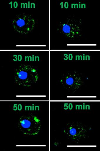
The team examined CHI3L1’s relationship with other key proteins and discovered why HPS patients can’t make use of CHI3L1 to prevent lung cells from dying. Without the HPS-1 mutation, CHI3L1 controls cell death by interacting with a protein on the surface of the cell called IL-13 receptor alpha2. It starts out inside the cell and traffics to the cell surface. But in mouse cells and human tissues with the HPS-1 mutation, the production of the receptor was decreased and its trafficking to the cell surface membrane was greatly diminished. The mutation prevents adequate amounts of IL-13 receptor alpha 2 from accumulating on the cell membrane to control cell death.
Toward therapies
In the mice, researchers stimulated cells to make much more of the IL-13 recepter alpha 2 protein than normal. With production of the protein in overdrive, enough finally accumulated in cell membranes of HPS-1 mice to keep more of the cells alive.
As they continued their research, the team pinpointed the protein that CHI3L1 works with to stimulate the collagen repairs. It’s called CRTH2. When they intervened to suppress CRTH2 function in HPS-1 mutant mice, they were able to slow down the scarring.
Elias said the research team has been in touch with companies that make drugs to suppress CRTH2. A new goal is to see if they can produce a possible life-extending therapy for HPS patients with pulmonary fibrosis.
“We have more work to do experimentally,” Elias said. “At that point, if we can show that the drugs that have already been produced by the different companies work in our model systems then taking them into man ought to move forward as quickly as possible.”
Meanwhile, finding ways to protect cells from dying using the insights of the study could be another therapeutic avenue.
In addition to Elias, Zhou, and Lee, the paper’s other authors are co-lead author Chuan Hua He, Erica Herzog, Xueyan Peng, Chang-Min Lee, Tung Nguyen, Mridu Gulati, Bernadette Gochuico, William Gahl, and Martin Slade.
The American Thoracic Society, the Hermanksy-Pudlak Syndrome Network and the National Institutes of Health funded the study (grants: HL-R01 HL093017, U01HL108638, R01HL-109033, R01HL115813, P20GM103652).

