PROVIDENCE, R.I. [Brown University] — Researchers from Brown University and the University of Rhode Island have demonstrated a promising new way to increase the effectiveness of radiation in killing cancer cells.
The approach involves gold nanoparticles tethered to acid-seeking compounds called pHLIPs. The pHLIPs (pH low-insertion peptides) home in on high acidity of malignant cells, delivering their nanoparticle passengers straight to the cells’ doorsteps. The nanoparticles then act as tiny antennas, focusing the energy of radiation in the area directly around the cancer cells.
In a paper published in the Proceedings of the National Academy of Sciences, the research team shows that the approach substantially increases the cancer-killing power of radiation in lab tests.
“This study was a good proof of concept,” said Michael Antosh, assistant professor (research) in Brown’s Institute for Brain and Neural Systems and the paper’s lead author. “We’re encouraged by our initial results and we’re excited to take the next step and test this in mice.”
The team is hopeful that the approach could ultimately improve radiation treatment for cancer patients. By increasing the effectiveness that a given dose of radiation has on cancer, the technique could reduce the overall radiation dose a patient requires, which would in turn reduce side effects. It could also increase the effectiveness of radiation at doses currently administered.
Special delivery
This research is an extension of work started by Yana Reshetnyak and Oleg Andreev, professors in the URI’s Division of Biological and Medical Physics, and professor Donald Engelman of Yale University, the inventors of pHLIP technology. The URI/Yale team had previously developed pHLIPs as a potential delivery system for cancer drugs and diagnostic agents. Cancer cells are generally more acidic than healthy cells, and pHLIPs are natural acid-seekers.
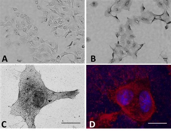
“We previously demonstrated that pHLIP-nanogold particles could find and accumulate in tumors established in mice,” Reshetnyak said. “Now our task is to test if we can treat cancer by irradiating tumors with nanogold particles more efficiently in comparison with traditional radiation treatment.”
Both theoretical and experimental work had shown that gold nanoparticles could intensify the effect of radiation. The particles absorb up to 100 times more radiation than tissue. Radiation causes the particles to release a stream of electrons into the area around them. If the particles were in close proximity to cancer cells, that stream of electrons would inflict damage on those cells.
“The idea here was to bring this all together, combining the nanoparticles with the delivery system and then irradiating them to see if it had the desired effect,” said Leon Cooper, the Thomas J. Watson Sr. Professor of Science at Brown and one of the study’s co-authors. Cooper, who shared the Nobel Prize in 1972 for explaining the behavior of electrons in superconductors, has been working for the last several years to better understand biological responses to radiation.
Auger effect
Gold is an especially good choice for amplifying radiation. When matter is hit by radiation at certain energies, electrons are released through a process known as the photoelectric effect. But gold has an additional source of electron emission, known as the Auger effect, that results from the particular arrangement of electrons orbiting gold atoms. It’s the effect of the Auger electrons that the researchers were working to maximize. Working out the quantitative details of the process involved complex calculations and simulations, Cooper said.
Auger electrons are low-energy and travel only a very short distance. Their travel distance is so short, in fact, that the electrons may not escape the nanoparticle if the particle is too large. So the researchers had to make sure their particles were small enough to emit those electrons. The short travel distance also means that particles need to be delivered in very close proximity to the cancer cells in order to do damage, hence the need for the pHLIPs.
Experiments showed that cancer cells irradiated in the presence of pHLIP-delivered gold had a 24-percent lower survival rate compared to those treated with radiation alone. The pHLIP samples had a 21-percent lower survival compared to irradiation with just gold but no pHLIPs. That suggests that the pHLIPs were effective in getting the gold close enough to the cells to do damage.
The next step, the researchers say, is to test the approach in a rodent model, which the team is planning to do soon.
“This work is a great example of successful collaboration between Brown and URI,” Andreev said. “We hope that the results of this research moving forward will lead to clinical application of pHLIP-based nanotechnology.”
Other authors on the study from URI were Dayanjali Wijesinghe, Samama Shrestha, and Natallia Katenka. Other authors from Brown were Robert Lanou, Yun Hu Huang, Nicola Neretti, Thomas Hasselbacker, David Fox, and Shouheng Sun. The work was supported by the National Institutes of Health (grants 2 P20 GM103430, CA133890, and GM073857).