PROVIDENCE, R.I. [Brown University] — Newly published research provides the first demonstration of how a genetic mutation associated with a common form of albinism leads to the lack of melanin pigments that characterizes the condition.
About 1 in 40,000 people worldwide have type 2 oculocutaneous albinism, which has symptoms of unsually light hair and skin coloration, vision problems, and reduced protection from sunlight-related skin or eye cancers. Scientists have known for about 20 years that the condition is linked to mutations in the gene that produces the OCA2 protein, but they hadn’t yet understood how the mutations lead to a melanin deficit.
In the new research a team led by Brown University biologists Nicholas Bellono and Elena Oancea shows that the protein is necessary for the proper functioning of an ion channel on the melanosome organelle, the little structure in a cell where melanin is made and stored. The ion channel is like a gate that lets electrically charged chloride molecules flow into and out of the melanosome. When the melanosome lacks OCA2 or contains OCA2 with an albinism-associated mutation, the researchers found, the chloride flow doesn’t occur and the melanosome fails to produce melanin, possibly because its acidity remains too high.
The discovery could inspire new ideas for treating albinism, said Elena Oancea, assistant professor of medical science and senior author of the paper published in the journal eLife.
“From a therapeutic point of view, we now have a channel that’s a possible drug target,” she said. Another potential treatment suggested by the research could be to alter melanosome acidity to make up for the lack of the protein.
A biology discovery
More generally, the study is also significant for being the first to show that ion channels are important for melanosomes to function properly. This wasn’t known before because melanosomes are generally too small for their electrical properties to be measured with the the technique of “patch clamping.” Such electrical readings are how biologists discover the comings and goings — the currents — of ions in cells, which is a fundamental process in cell physiology.
“I think it is a big step forward because not only did we make progress in understanding the function of one protein important in pigmentation, but we kind of opened up a new way to study how the melanosome operates,” said Bellono, a graduate student and the paper’s lead author. “There hasn’t been much research on ion channels in the melanosome.”
Because melanosomes are so small, Oancea and Bellono had to begin their study of the OCA2 protein and its mutant forms in organelle cousins of the melanosome, such as the endolysosome, because those can be made large enough for patch clamping. In experiments where they made endolysosomes express OCA2, for example, they measured currents related to the passing of chloride ions. That provided their first key evidence that the protein was associated with an ion channel.
They also used endolysosomes to find that that the OCA2 mutation V443I specifically affects the ion channel. That mutation decreased the chloride ion current by 85 percent compared to normal versions of the protein.
In another experiment Oancea and Bellono showed that expression of normal OCA2 in the endolysosomes, which are acidic organelles, reduced acidity to above 6 on the pH scale, which is required in a melanosome for the protein tyrosinase to trigger melanin production.
Into the melanosome
But to truly understand the role of OCA2 and the V443I mutation in albinism, the researchers needed to look directly at melanosomes. They were able to turn to helpful colleagues. Co-author Michael Marks at the University of Pennsylvania introduced them to a line of mutant mouse skin cells that had unusually large melanosomes. Anita Zimmerman, professor of medical science who works down the hall at Brown University, tipped them off that bullfrogs happen to have especially large melanosomes in their retinas.
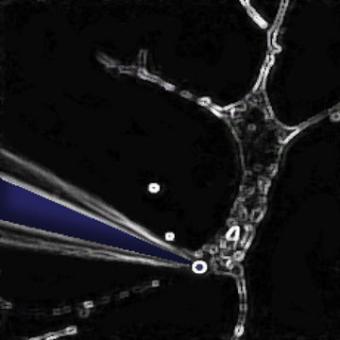
Patch clamp experiments with those large melanosomes confirmed the role of the V443I mutation in the failure of chloride ion channels. First, they compared chloride currents in normal melanosomes and ones in which they used interference RNA (a method of blocking gene expression) targeted to prevent OCA2 production. They found that the melanosomes without OCA2 produced much less current and much less melanin. Then they added either normal OCA2 or OCA2 with the V443I mutant, and they found that only the normal OCA2 protein could restore current and melanin production.
They did another experiment to ensure that melanin itself wasn’t responsible for the chloride current. It wasn’t.
Many details of the OCA2 protein’s role in the melanosome’s ion channels are still not known, the authors said, but the research points to the key mechanism that breaks down when it fails.
“OCA2 activity modulates the melanin content of melanosomes, most likely by regulating organellar pH,” they wrote in eLife. “We propose that OCA2 contributes to a novel melanosome-specific anion current that modulates melanosomal pH for optimal tyrosinase activity required for melanogenesis.”
In addition to Bellono, Oancea, and Marks, the paper’s other authors are Iliana Escobar of Brown and Ariel Lefkovith of Penn.
The National Institutes of Health (National Institute of Arthritis and Musculoskeletal and Skin Diseases, National Eye Institute and the National Institute of General Medical Scienes), the National Science Foundation, and Brown University provided funding for the study.

