PROVIDENCE, R.I. [Brown University] — A team of researchers has captured the dynamics of how a ring-shaped molecule opens and unfurls in an ultra-high speed “molecular movie.” This is the first time a chemical reaction has been imaged at the molecular level with such short time scales. The research could serve as a basis for understanding similar reactions involved in a wide variety of chemical processes.
The work, which was done at SLAC National Accelerator Laboratory in California, is described in a paper published in Physical Review Letters. Peter Weber, professor of chemistry and dean of Brown’s Graduate School, and graduate student James Budarz were co-authors of the new research.
Budarz spoke with Kevin Stacey about the work.
What exactly was the molecular process that you were able to image?
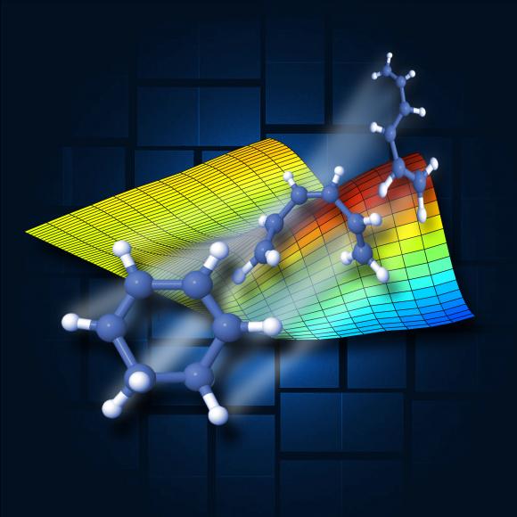
This experiment captured images of gas phase 1,3-cyclohexadiene as it underwent a ring-opening reaction to produce 1,3,5-hexatriene. Essentially we showed the step-by-step process by which the ring structure is broken open when it interacts with light.
The reaction has importance as a prototypical system. It has been studied for years as a fundamental stepping-stone to larger molecules. Despite a large body of work, including spectroscopic experiments that show the excited state potential energy surfaces in this molecule performed by our own research group at Brown, no technique had yet imaged the full structure during the reaction. Using ultrafast X-rays produced by a free-electron laser we’ve now been able to make a molecular movie of the process.
How was the imaging done?
The only place these X-ray experiments can be done is at the Linac Coherent Light Source at the SLAC Linear Accelerator Center in California. The light source is an X-ray free electron laser at the end of a two-mile long linear accelerator that produces short, bright bursts of X-ray photons.
One of the problems in capturing this reaction is that it happens so fast. The reaction is complete within 200 femtoseconds — 2x10-13 seconds. LCLS is the first X-ray source with sufficient brightness and short enough pulses to image the reaction before it’s over.
We used what’s known as a pump-probe scheme. The reaction is triggered by a pulse of ultraviolet laser light, which is followed closely by the X-ray pulse. When the X-ray pulse strikes the molecule, some of the photons are diffracted and then collected by an imaging detector that records their distribution in space. By varying when the X-ray arrives and assembling a sequence of these images, the frames become a movie and the transient structures are captured.
Why is it useful to know how the reaction works?
Ring structures in molecules are abundant in nature, as are light-induced reactions similar to the one we imaged. For example, the formation of pre-vitamin D in the skin has a ring-closing reaction upon UV excitation, which is similar to the reaction we studied.
More importantly, the success of this experiment opens doors for many more studies of chemical dynamics in more complex systems.
What’s the next step in this work?
We’ve been awarded more beam time to perform another experiment coming up in 2016. With this opportunity, we hope to expand our technique to explore more classes of photo-induced reactions.